3 Ligaments Of The Lateral Ankle / Broken Ankle Vs Sprain Symptoms And Recovery Time / They run from the lateral malleolus of the fibula to the talus and calcaneus.
Get link
Facebook
X
Pinterest
Email
Other Apps
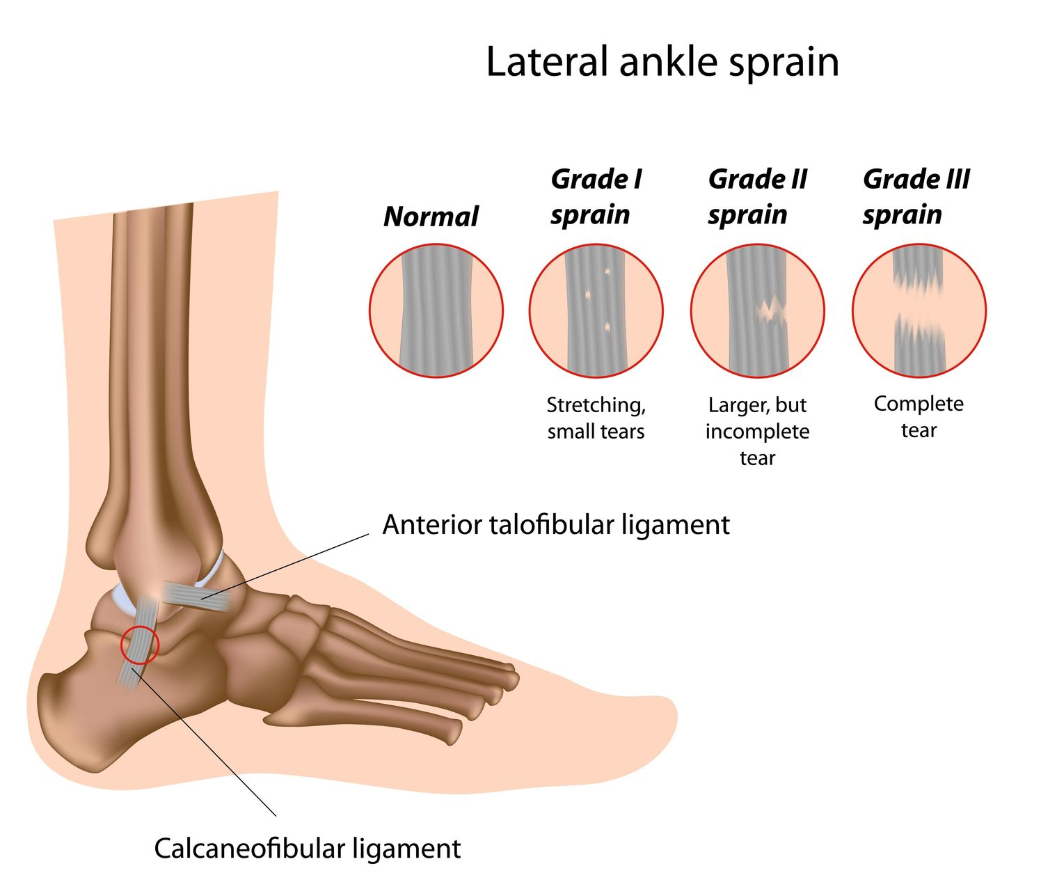
3 Ligaments Of The Lateral Ankle / Broken Ankle Vs Sprain Symptoms And Recovery Time / They run from the lateral malleolus of the fibula to the talus and calcaneus.. The lateral ankle ligament is a complex of three different ligaments including the calcaneofibular ligament (cfl), anterior talofibular ligament (atfl), and the posterior talofibular ligament (ptfl). The posterior tfl is rarely disrupted from inversion injury, unless such injury is accompanied by ankle dislocation ( , 3 ). The anterior tibiofibular ligament (2), which connects the tibia to the fibula; Full weight bearing in cast boot. The job of a ligament is to join bone to bone and provide stability to a joint.
Sprains can range from tiny tears in the fibers that make up the ligament to complete tears through the tissue. Three ligaments on the outside of the ankle that make up the lateral ligament complex, as follows: Ankle brace when out of bed until balance returns They are more commonly injured than the medial collateral (deltoid) ligament of the ankle. The anterior talofibular ligament (atfl), which connects the front of the talus bone to a long bone in the lower leg called the fibula the calcaneofibular ligament (cfl), which connects the calcaneus, or heel bone, to the fibula
Sprained Ankle Definition Anatomy And Causes Video Town Center Orthopaedic Associates from jeffreybergmd.com The lateral collateral ligament consists of three ligaments: In this video we are going to learn how the injuries of lateral ankle ligaments are evaluated with ultrasound. Remove for showers after md removes surgical dressing. The posterior tfl is rarely disrupted from inversion injury, unless such injury is accompanied by ankle dislocation ( , 3 ). These three ligaments work together to prevent the ankle from inversion. If there is a complete tear of the ligaments, the ankle may become unstable after the initial injury phase passes. They run from the lateral malleolus of the fibula to the talus and calcaneus. Wear day & night, remove for showers only.
The anterior talofibular ligament (atfl), the calcaneofibular ligament (cfl), and the posterior talofibular ligament (ptfl;
And, on the medial side of the ankle, the deltoid ligaments (4), which connect the tibia to the talus and. The lateral collateral ligaments (3), which attach the fibula to the calcaneus and gives the ankle lateral stability; Full weight bearing in cast boot. Complications of ankle sprains can also cause lateral ankle pain sometime after the injury has. Kobayashi t(1), suzuki d(2), kondo y(3), tokita r(4), katayose m(5), matsumura h(5), fujimiya m(6). If there is a complete tear of the ligaments, the ankle may become unstable after the initial injury phase passes. 3 major lateral ankle ligaments: Three ligaments on the outside of the ankle that make up the lateral ligament complex, as follows: In this video we are going to learn how the injuries of lateral ankle ligaments are evaluated with ultrasound. They run from the lateral malleolus of the fibula to the talus and calcaneus. Most sprained ankles occur in the lateral ligaments on the outside of the ankle. They are more commonly injured than the medial collateral (deltoid) ligament of the ankle. The anterior talofibular ligament (atfl), which connects the front of the talus bone to a long bone in the lower leg called the fibula the calcaneofibular ligament (cfl), which connects the calcaneus, or heel bone, to the fibula
If there is a complete tear of the ligaments, the ankle may become unstable after the initial injury phase passes. The anterior talofibular ligament (atfl), which connects the front of the talus bone to a long bone in the lower leg called the fibula the calcaneofibular ligament (cfl), which connects the calcaneus, or heel bone, to the fibula Most sprained ankles occur in the lateral ligaments on the outside of the ankle. The lateral collateral ligament consists of three ligaments: The lateral collateral ligaments (3), which attach the fibula to the calcaneus and gives the ankle lateral stability;
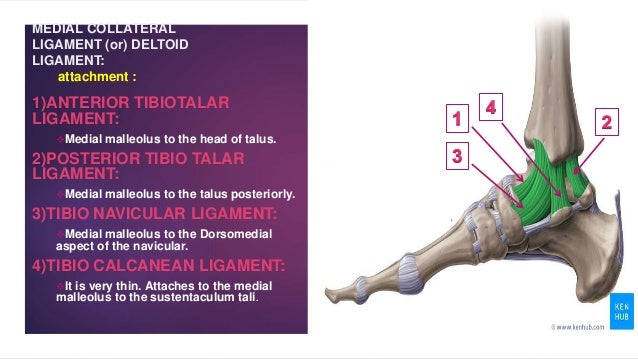
Ligaments Of Ankle Joint Ankle Complex from image.slidesharecdn.com What is a lateral ankle ligament injury? These three ligaments work together to prevent the ankle from inversion. The lateral collateral ligaments (3), which attach the fibula to the calcaneus and gives the ankle lateral stability; Lateral ankle ligament reconstruction rehabilitation protocol weight bearing: And, on the medial side of the ankle, the deltoid ligaments (4), which connect the tibia to the talus and. They run from the lateral malleolus of the fibula to the talus and calcaneus. The three medial collateral ligaments are better known as the deltoid ligament. The lateral collateral ligament (complex) of the ankle is a set of three ligaments that resist inversion of the ankle joint.
The lateral collateral ligament (complex) of the ankle is a set of three ligaments that resist inversion of the ankle joint.
Connects talus to fibula thickening of a capsule weakest link prevents anterior sublux check with anterior drawer. The most common ankle injury is a sprained ankle. And, on the medial side of the ankle, the deltoid ligaments (4), which connect the tibia to the talus and. The major ligaments of the lateral ankle consist of the anterior talofibular ligament, the posterior talofibular ligament, and the calcaneal fibular ligament. If you have suffered an inversion ankle sprain it means you have injured one or more of the three main ligaments on the outside of your ankle; The three medial collateral ligaments are better known as the deltoid ligament. 3 major lateral ankle ligaments: In this video we are going to learn how the injuries of lateral ankle ligaments are evaluated with ultrasound. The anterior talofibular ligament, the posterior talofibular ligament, and the calcaneofibular ligament. Ankle brace when out of bed until balance returns An ankle sprain usually happens when the foot inverts or rolls out, stretching or tearing the ligaments and tendons on the outside of the ankle. Ankle sprains most often involve injuries to the ligaments that stabilize the lateral ankle. The anterior talofibular ligament (atfl), the posterior talofibular ligament (ptfl) and the calcaneofibular ligament (cfl).
The lateral collateral ligaments (3), which attach the fibula to the calcaneus and gives the ankle lateral stability; Lateral ankle ligament reconstruction rehabilitation protocol weight bearing: The anterior tibiofibular ligament (2), which connects the tibia to the fibula; Chronic ankle instability following ankle sprains causes pain and functional problems such as recurrent giving way. In this video we are going to learn how the injuries of lateral ankle ligaments are evaluated with ultrasound.
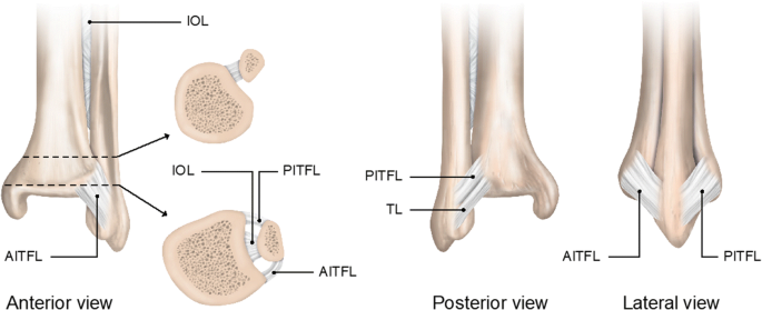
Return To Play After A Lateral Ligament Ankle Sprain Springerlink from media.springernature.com The lateral collateral ligament consists of three ligaments: Important ligaments of the ankle are those that compose the distal tibiofibular syndesmosis and the lateral and medial collateral ligaments (, 1,, 2). An ankle sprain usually happens when the foot inverts or rolls out, stretching or tearing the ligaments and tendons on the outside of the ankle. Most sprained ankles occur in the lateral ligaments on the outside of the ankle. Wear day & night, remove for showers only. The anterior talofibular ligament, the posterior talofibular ligament, and the calcaneofibular ligament. This ligament protects against inversion & plantarflexion. Anterior talofibular calcaneofibular posterior talofibular.
The most common ankle injury is a sprained ankle.
The medial (deltoid) ligaments is much stronger than the lateral ligament and is therefore injured much less frequently. The major ligaments of the ankle are: If you have suffered an inversion ankle sprain it means you have injured one or more of the three main ligaments on the outside of your ankle; The posterior tfl is rarely disrupted from inversion injury, unless such injury is accompanied by ankle dislocation ( , 3 ). Within the 3 ligaments of the lateral ligament complex, 80% of patients tear the anterior talofibular ligament (atfl), whereas the other 20% of patients tear the atfl and calcaneofibular ligament (cfl). 574.247.9442 www.sbortho.com lateral ankle sprain nonoperative protocol page 1 of 2 last updated september 3, 2020 lateral ankle sprain nonoperative protocol ankle sprains (stretching of the lateral ankle ligaments) are one of the most common injuries to occur in the lower extremity. This ligament protects against inversion & plantarflexion. Chronic ankle instability following ankle sprains causes pain and functional problems such as recurrent giving way. Lateral ankle ligament injury is a common msk condition representing 85% of ankle injuries and has a high recurrence rate (up to 70%). Sprains can range from tiny tears in the fibers that make up the ligament to complete tears through the tissue. Important ligaments of the ankle are those that compose the distal tibiofibular syndesmosis and the lateral and medial collateral ligaments (, 1,, 2). The anterior talofibular ligament (atfl), which connects the front of the talus bone to a long bone in the lower leg called the fibula the calcaneofibular ligament (cfl), which connects the calcaneus, or heel bone, to the fibula Remove for showers after md removes surgical dressing.
They are more commonly injured than the medial collateral (deltoid) ligament of the ankle 3. liga. The many ligaments of the ankle complex hold 14 bones in close proximity, including the tibia, fibula, and tarsals.
Comments
Post a Comment